| Chapter 4: | Examination of the Chest | |
| Page 3 0f 4 |
 |
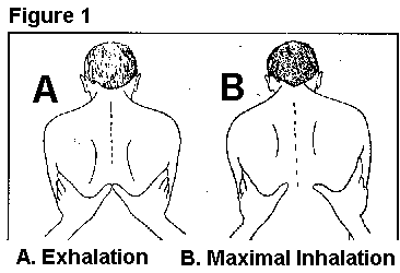
Thoracic expansion The patients chest wall normally expands symmetrically during deep inhalations, and the RCP can evaluate the expansion on both the anterior and posterior chest. The RCP first places hands over the anterolateral chest, extending the thumbs along the costal margin toward the xiphoid. To evaluate it posteriorally, the hands are placed over the posterolateral chest, with thumbs joining at approximately T8. Instruct the patient to exhale slowly and completely. Once completed, the RCPs fingertips are secured against the sides of the patients chest, extending the thumbs toward the midline until their tips meet at the midline. The patient is then instructed to take a full deep breath. The RCP takes note of the distance each thumb moves away from the midline. Normal movement is about 3-5 cm for each thumb. |
|
Bilateral reduction in chest expansion can usually be seen in patients with diseases affecting the expansion of both lungs. Unilateral reduction in expansion is indicative of a respiratory disease that impedes expansion of one lung, such as lobar consolidation, pleural effusion, atelectasis, or pneumothorax.
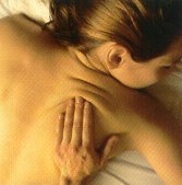
The skin and subcutaneous tissues of the chest wall should be palpated to ascertain the general temperature and condition of the skin. When there is air leaking from the lung into the subcutaneous tissues, fine bubbles create crackling sounds and sensations when being palpated. This condition is called subcutaneous emphysema, and the sensation produced during palpation is referred to as crepitus.
Percussion
The act of tapping on a surface in order to evaluate the underlying sound is called percussion, and percussion of the patients chest wall creates a sound and palpable vibration that is useful in evaluating underlying lung tissue. The vibration created by percussion penetrates the lung to a depth of about 5-7 cm below the chest wall.
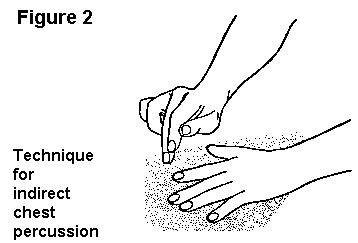
To perform chest percussion, the middle finger of the left hand is placed firmly on the area to be percussed. The back of it middle phalanx is then struck with the tip of the middle finger of the right hand. Deliver the stroke from the wrist and finger joints, bending the percussing finger so its terminal phalanx is at right angles to the metacarpal bones when the blow is delivered, and it strikes the pleximeter finger in a perpendicular way.
|
Percussion over normal lung fields produces a low-pitch sound that is easy to hear (referred to as normal resonance). Resonance is said to be increased when the percussion note is louder and lower in pitch. Percussion may also produce a high-pitched, short in duration sound that is dull or flat, just the opposite of resonance. The percussion of the chest alone has little clinical implication. However, when it is considered with other findings, it can yield essential information.
Chest percussion is generally performed evaluate the extent of diaphragmatic excursion and air-fluid levels. The note heard on percussion becomes more resonant as the diaphragm descends and lungs fill with air. When the sound changes to a dull note, it indicates the limit of diaphragm descent. The less resonant the percussion notes are indicative of tissues that are more dense. As a result, air naturally produces the most resonant notes, such as is heard over a pneumothorax. Normal lung tissue produces duller notes, with accumulation of fluids producing even duller notes. The dullest of all notes are heard when percussing over bone structure.
Abnormalities that increase lung tissue density, such as atelectasis or pneumonic consolidation, result in a loss of resonance and a dull percussion note above the affected area.