| Chapter 5: | Analysis of Chest X-rays | |
| Page 1 0f 1 |
As a result of the patient interview and examination, the RCP can form some initial conclusions regarding a diagnosis of the patientís condition. These conclusions will prove valuable in helping the RCP assure that the therapeutic modalities prescribed by the physician are best suited for the patient.
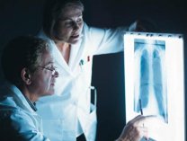
| In addition to interviewing and examining patients, RCPs need to be able to evaluate a patientís chest x-rays. This module is not intended to provide you with the interpretive skills of a radiologic technologist. It is designed to enable you to recognize structures and basic areas of normality and abnormality. |  |
Diagnostic radiographs of the chest are an important part of the evaluation of the patient with respiratory complaints.
In a chest radiograph, you can see the patientís heart left-center, and the left ventricle will appear to be most prominent. The other features that can be observed include:
- Aortic knob: It lies superior to the heart, and can be distinguished by its rounded appearance.
- Right atrium: Along with the superior vena cava, it appears to be the heartís border on the right side.
- Descending aorta: Appears posteriorly to the heart in lateral view x-rays.
- Lungs: Appear on both side of the chest, and have the look of translucent shadows.
- Diaphragm: This dome-shaped structure can be seen located along the inferior border of the chestís cavity. Its right side is slightly (2 cm) higher because of the location of the liver.
- Hilium: Can be seen in the medial area of the chest, and its pulmonary vessels and lymph nodes appear to be a branching density.
X-rays also reveal the presence of infiltrates, which are usually caused by blood or body fluids accumulating in the vascular space. These appear as darkened or clouded areas on the radiographs. When the alveoli fill with fluids or the tissues consolidate around the bronchus, that area will be clearly visible on the radiograph. If a density or clouded area appears to be anterior to the heart, the border will be obscured. Densities lying posteriorly to the heart do not obscure the heartís border.
The most common and useful chest x-rays are the posterior-anterior (PA) views. However, bedside radiographs sometimes are required as a result of the patientís condition. These portable x-rays donít allow as much control of positioning or film exposure, and in AP films, the heart usually appears larger because it is a greater distance from the film plate.
Some of the other radiographic procedures that can facilitate diagnosis of a patient include:
- Fluoroscopy: This technique permits the heart and lungs can be viewed in motion. It facilitates the diagnosis of paralysis of any part of the diaphragm since it can be observed moving during the patientís breathing process.
- Inspiration/Expiration Films: These radiographs are taken as the patient breaths maximally in and out. They can help confirm the presence of bronchial obstructions, including tumors or blebs frequently seen in COPD patients.
- Oblique views: Taken at approximately a 45° angle to the film plate, these chest views allow a review of the heartís more difficult to see areas, and facilitate assessing its size. Effective viewing of the lung apices is permitted by the lordotic view, which involves lifting the clavicles away from the lung tissue.
- Bronchoscopy: This technique involves insertion of a fiberoptic endoscope into the bronchi, and has become most valuable for diagnosing the presence of lung cancer.
- Computed tomography (CT): This technique utilizes computer technology to evaluate "slices" of lung density data. It is valuable and effective for: observing focal lung lesions beneath bony areas, identifying the bullae and blebs seen in pulmonary emphysema, and can help physicians analyze the lesions seen in lung cancer prior to performing surgery.
- Magnetic resonance imaging (MRI): This relatively new non-invasive painless technique involving electromagnetic field technology has proven to be valuable in evaluating the possibility of cancer in the chest wall and mediastinum, and has proven useful for diagnosing thoracic aneurysms, congenital anomalies and major vessels of the aorta.