| Chapter 3: | Physical Examination | |
| Page 1 0f 4 |
Introduction
The patient interview and the RCPs initial observations yield a great deal of valuable assessment information. However, the actual physical examination of the pulmonary patient is most valuable in facilitate the RCPs accurate evaluation of the patients condition and subsequent prescription of a treatment protocol.
Vital Signs
| The RCPs hands-on physical examination includes checking the patients vital signs. | 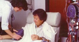 |
While parts of the following discussion may seem somewhat basic, it is important that your review of patient assessment skills needed to develop and implement therapist driven protocols include a brush-up on what you know likely already know about conducting physical evaluations of pulmonary patients.
The vital signs are a nonspecific but necessary part of any physical examination, and assessment of the vital signs is the most frequent evaluation technique performed in the clinical setting. The patients vital signs provide crucial information and clues regarding the patients overall health status, and their response to treatments.
Many times during a physical examination, the measuring the vital signs often gives initial evidence of an abnormality. The four basic vital signs are body temperature, pulse rate, blood pressure and respiratory rate. While an in-depth discussion of the vital signs is beyond the scope of this CEU, checking of vital signs should always be considered as part of a patient assessment:
I. Heartbeat
To begin the assessment of vital signs, the RCP needs to be adept at taking the patients pulse. A pulse indicates a heartbeat and can be felt at any of the patients arteries. Documentation of the patients pulse should include the frequency, regularity, and quality of the heartbeat. Pulses monitored in adults include the radial, carotid, or femoral pulses. In children and infants, the brachial pulse is preferred. In the documentation process, it is important to note the rate per minute, as well as the regularity and quality of the pulse.
The amount of oxygen being delivered to the patients tissues is dependent on the hearts ability to pump oxygenated blood through the circulation system. The amount pumped per minute, cardiac output, is a direct function of heart rate and stroke volume. When the oxygen content of arteries dips below normal, often as a result of lung disease, the patients heart attempts to maintain normal oxygen delivery by increasing the cardiac output. This is achieved by increasing the heart rate.
The patients radial artery is most commonly used to assess the pulse rate. The number of times the heart beats per minute is measured by counting the pulse in the artery. The RCP places the second and third finger pads on the radial pulse to count for about one minute. Be careful not to hold the patients wrist too far above the heart because that can make obtaining an accurate pulse difficult. The normal range for adult heart rates is between 60-100 beats per minute (bpm). The average adult pulse rate is 72/bpm.
A heart rate slower than 60/bpm is called bradycardia, while tachycardia is a rate greater than 100/bpm. A normal pulse beats in consistent intervals, and when the interval varies from beat to beat, the pulse is considered to be irregular.
The pulse rate is influenced by several factors, with exercise being the most obvious. With increased activity, the heartbeat increases 20-30 beats per minute to meet the bodys needs. It should return to normal within 3 minutes after the activity has ceased. The heart rate also increases in response to fear, anxiety, low blood pressure, anemia, fever, hypoxia, some medications and for many other reasons. Heart rate decreases with hypothermia, certain arrhythmias, due to medications and other reasons.
Remember that spontaneous ventilation can influence pulse strength (amplitude) changes. A significant decrease in pulse amplitude during inhalation is known as pulsus paradoxus (paradoxical pulse). This is common in patients afflicted with obstructive disease, particularly those experiencing an acute asthma attack. Pulsus paradoxus also signals the possible existence of mechanical restriction of the hearts pumping action, such as is seen in constrictive pericarditis or cardiac tamponade. Taking a blood pressure measurement best assesses this condition. An alternating succession of strong and weak pulses, pulsus alterans, suggests left- sided heart failure and is not related to the presence of any respiratory diseases.
Evaluating the carotid, femoral, brachial, temporal, popliteal, posterior tibial, and dorsalis pedis can also assess the patients pulse. The carotid and femoral pulse should be used when the blood pressure is abnormally low. To find the carotid pulse, locate the larynx with the tips of your first two or three fingers, slide your fingers away from the larynx (Adams apple) towards the groove between the trachea and the large neck muscles, and feel for the pulse. Move your fingertips around until you find the strongest point and feel the pulse. Never use your thumb because it has a pulse of its own and could be mistaken for the patients pulse. Count the pulse rate and note whether it is strong, weak, regular or irregular.
If the carotid site is used, you should take care to avoid the carotid sinus area because it can evoke a strong parasympathetic response, causing bradycardia or asystole. To obtain a femoral pulse, visualize the crease between the leg and the abdomen, place the tips of your first two or three fingers at the midpoint, and feel for the pulse.