| Chapter 3: | Physical Examination | |
| Page 2 0f 4 |
II. Blood Pressure
Blood pressure is an indication of how well the heart is pumping, how much blood it pumps, and how efficiently the job is performed. The pressure is the pressure of the blood against the walls of the blood vessels.
The force exerted on the walls of the arteries as blood pulses through them is called the arterial blood pressure. Arterial systolic blood pressure represents the peak force that is exerted during the contraction of the hearts left ventricle. Diastolic pressure indicates the force that remains after relaxation. Pulse pressure is the variance between systolic and diastolic pressures. For example, if systolic pressure is 120 and diastolic pressure is 100, the pulse pressure is 20. Normal pulse pressure ranges between 35-40 mm Hg. When the pulse pressure measures less than 30 mm Hg, peripheral pulse is difficult to detect.
On the other hand, the patients blood pressure is determined by: the force of the left ventricular contraction, the systemic vascular resistance, and the blood volume. Normal systolic pressure ranges from 95-140 mm Hg, with an average of about 120 mm Hg. Normal diastolic pressure ranges from about 60-90 mm Hg, with the average norm being 80 mm Hg. Blood pressure is recorded as a fraction, by listing the systolic pressure over the diastolic pressure. For example, normal blood pressure is recorded as being 120/80 mm Hg.
Blood pressure rises or drops for a variety of reasons. Hypotension is defined as a drop in blood pressure to a measurement of less than 95/60 mm Hg. The most common causes of hypotension include: ventricular failure, peripheral vasodilatation, or low blood volume. Vital body organs receive inadequate blood flow in patients with hypotension. Inadequate circulation can impair O2 delivery to the tissues, causing tissue hypoxia to occur. As a result, it is important that prolonged hypotension be prevented.
When persons with normal blood pressure sit or stand up, blood pressure is relatively unaffected. However, similar actions may cause an abrupt drop in blood pressure among hypovolemic patients. This condition, which is referred to as postural hypotension, can be confirmed by measure the patients blood pressure in both the supine and sitting positions.
Sudden decreases in arterial blood pressure caused by postural hypotension can decrease cerebral blood flow, leading to syncope, or fainting. Treatment of postural hypotension involves administration of fluid or vasoactive drugs. It is important that untreated or unresponsive postural postural hypotension be considered when moving or ambulating a patient.
The systolic blood pressure generally decreases slightly with normal inhalation; however, a drop of more than 6-8 mm Hg during resting inhalation is abnormal and referred to as paradoxical pulse. Paradoxical pulse is caused by intrathoracic pressure swings created by the respiratory muscles during breathing. Negative intrathoracic pressure during inspiration assists venous return to the right ventricle (RV), however it impedes arterial outflow from the left ventricle (LV). Increased venous return increases RV pressures, thus restricting LV filling. This in turn reduces LV stroke volume and decreases systolic blood pressure during inhalation. Palpation may indicate the presence of paradoxical pulse, but it can only be quantified by ausculatory measurement.
Increases in patient blood pressure can have even more serious consequences. Blood pressure that consistently measures above 140/90 mm Hg is referred to as hypertension, which is usually caused by high systemic vascular resistance. A less common cause of hypertension is the increased force of ventricular contraction. Hypertension that is severe often results in congestive heart failure, central nervous system abnormalities, uremia, or cerebral hemorrhage leading to the patient suffering a stroke.
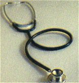 |
Measuring the patients blood pressure is most commonly accomplished the auscultatory method. This involves the use of a stethoscope and a sphygmomanometer. Be sure to check the stethoscope to be sure its in good working order prior to use. The sphygmomanometer measures arterial blood pressure, and consists of a manometer containing a scale for registering pressure as well as an inflatable bag surrounded by a covering known as a cuff. |
Remember that cuffs come in various sizes, and that it is crucial that the proper-size cuff be used. Using the wrong-size cuff can cause erroneous blood pressure readings to be obtained. For example, if the cuff is too narrow for the patients upper arm, the reading obtained will be falsely high. A regular-sized cuff should be used if the arms circumference is less than 13 inches, arm greater than a 13 inch circumference require use of a large-size cuff, and pediatric patients or adults with extremely small arms require use of a pediatric cuff.
When the RCP applies the cuff to the patients upper arm and pressurizes it to exceed systolic blood pressure, brachial blood flow is stopped. As the cuffs pressure is decreased slowly (at a rate of about 2-3 mm Hg per second) to the point just below systolic pressure, blood flow intermittently passes the obstruction. This partial obstruction of blood flow creates vibrations and turbulence called Korotkoff sounds, which can be heard by placing the stethoscope over the brachial artery distal to the obstruction.
At the point at which the first Korotkoff sounds are heard, systolic blood pressure is recorded. The point at which the sounds become muffled is the diastolic pressure, and this muffling is the final change in the Korotkoff sounds prior to their disappearance. If the muffling and cessation of the sounds occur at a wide interval, all three pressures should be recorded (120/80/60).
Besides using the wrong-sized cuff, other mistakes that cause erroneously high blood pressure measurements include:
- Cuff applied either too tightly or too loosely
- Excessive pressure placed in the cuff during measurement
- Inflation pressure held in the cuff
- Incomplete deflation of cuff between measurements
Extraneous room sounds, ventilators, static electricity, or the presence of an auscultatory gap may also cause errors in measurement of the patients blood pressure. The auscultatory gap consists of a 20-40 mm Hg drop with no sound between the first systolic sound and the continuous pulse sound. Inflating the cuff until the palpated radial pulse can no longer be felt can help prevent missing the opening snap. When the auscultatory gap is heard, both the opening snap pressure and pressure at which continuous pulses are heard should be recorded (160/140/80).